")
")
| Issue |
Radioprotection
Volume 59, Number 4, October - December 2024
|
|
|---|---|---|
| Page(s) | 278 - 286 | |
| DOI | https://doi.org/10.1051/radiopro/2024025 | |
| Published online | 13 December 2024 | |
Article
Evaluation of barium sulfate-copper breast radiation shield for use in thoracic computed tomography examinations
1
Medical Physics Department, Faculty of Medicine, Semnan University of Medical Sciences, Semnan, Iran
2
Cancer Research Center, Semnan University of Medical Sciences, Semnan, Iran
3
Medical Physics and Radiology Department, Faculty of Medicine, Infectious Diseases Research Center, Gonabad University of Medical Sciences, Gonabad, Iran
4
Biomedical Engineering and Medical Physics Department, School of Medicine, Shahid-Beheshti University of Medical Sciences, Tehran, Iran
5
Social Determinants of Health Research Center, Semnan University of Medical Sciences, Semnan, Iran
* Corresponding authors: This email address is being protected from spambots. You need JavaScript enabled to view it.
; This email address is being protected from spambots. You need JavaScript enabled to view it.
Received:
5
May
2024
Accepted:
4
July
2024
Abstract
Introduction: In thoracic computed tomography (CT) examinations, patients’ breasts are exposed to high radiation doses, necessitating the reduction of received dose by a radiation shield. The aim of this study is to evaluate the efficiency of a new composition of barium sulfate-copper shield with minimal impact on image quality. Materials and methods: Different breast shields were manufactured using varying weight percentages of copper and BaSO4. Thermoluminescent dosimeters (TLDs) and thorax phantoms were employed to assess the radiation shielding effectiveness. Image quality, in terms of noise and CT number accuracy, was quantitatively evaluated on a CT dose Index phantom (CTDI). Additionally, a controlled trial involving with 30 female participants was conducted to further assess CT image quality and select the best breast radiation shield. Results: The results indicated that the different shield compositions reduced the surface dose by 14.17–51.69%. The shield with a composition of 90% Cu–10% BaSO4 and 50% Cu–50% BaSO4 had the lowest noise, while the 100% bismuth shield had the highest noise. Importantly, the 50% Cu–50% BaSO4 shield did not cause artifacts in the thoracic CT images. Conclusion: By using the 50% Cu–50% BaSO4 shield, a significant dose reduction was achieved while maintaining appropriate image quality, making it suitable for clinical applications.
Key words: Dose reduction / breast radiation shield / thorax CT scanning / image quality / image artifact
© F. Poursoltani et al., Published by EDP Sciences, 2024
 This is an Open Access article distributed under the terms of the Creative Commons Attribution License (https://creativecommons.org/licenses/by/4.0), which permits unrestricted use, distribution, and reproduction in any medium, provided the original work is properly cited.
1 Introduction
The use of computed tomography (CT) as a diagnostic imaging technology has increased in the last two decades (Abolhadi et al., 2023; Bertho et al., 2024; Lestari et al., 2023). Compared to the other imaging modalities, CT examinations use ionizing radiation with higher doses (Curtis, 2010; Saba et al., 2019; Schöckel et al., 2020), contribute to 70% of the overall dose of medical patients (Saba et al., 2019). A report in 2005 indicated that CT scan accounted for only 11% of all radiological tests (Curtis, 2010). Today, the number of CT examinations around the world increases by 4% annually, which is a total of about 300 million CT scans per year (Schöckel et al., 2020).
CT scans are essential examinations for efficiently diagnosing a wide variety of thoracic diseases and are the most accurate technique for lung examinations (Alonso et al., 2016; Saba et al., 2019). Exposure of radiation-sensitive organs can lead to an increased risk of cancer (Alonso et al., 2016; Dere, 2022; Liao et al., 2019). The cancer risk from chest CT is estimated to be between 25 and 33 cases per 100,000 examinations (Colletti et al., 2013). In thoracic CT examinations, the breast tissue, as a radiation-sensitive organ, receives a large dose due to its superficiality and its location in the scanning field, even though it is not under clinical examination (Lestari et al., 2023; Mehnati et al., 2023; Saba et al., 2019). In chest CT, the dose delivered to the breast is usually 0.035–0.02 Gy (Saba et al., 2019; Tappouni et al., 2013). Due to the breast’s high radiation sensitivity, with a weighting factor of 0.12, and its exposure to high doses during chest CT examinations, reducing the breast dose is important (Lestari et al., 2023; Saba et al., 2019).
There are several techniques to reduce breast dose during thoracic CT examinations: (1) Iterative Reconstruction Algorithm (IRA) (Foley et al., 2013; Lambert et al., 2016); (2) Tube Current Modulation (TCM) techniques (angular or organ-based) (Feldle et al., 2023; Foley et al., 2013; Lambert et al., 2016; Saba et al., 2019); and (3) Using bismuth shield (Mehnati et al., 2019; Mehnati et al., 2019; Saba et al., 2019). The use of a bismuth shield is an effective method for reducing breast absorbed dose during CT examinations. By absorbing low-energy photons, the bismuth shield reduces the dose to the superficial organs by 26–57%. Bismuth breast shields work independently of the CT scanner and can therefore be used with scanners of different models and manufacturers. The advantages of bismuth breast shields include low cost, effectiveness, and ease of manipulation (Ko et al., 2021). Despite the benefits of dose reduction, the bismuth shield has a detrimental effect on image quality, such as changes in the accuracy of the CT number and an increase in image noise and artifacts (Foley et al., 2013; Lambert et al., 2016; Liao et al., 2019; Saba et al., 2019). The other researchers (Alonso et al., 2016; Colletti et al., 2013; Einstein et al., 2012; Huggett et al., 2013) have conducted further investigations on the use of bismuth shields. They demonstrated that the use of bismuth shield reduced the breast surface dose but it also increased image noise, created streak artifacts and decreased image quality (signal and contrast). The usefulness of bismuth shield has been challenged by the American Association of Physicists in Medicine (AAPM) (AAPM Report, 2024; Akhlaghi et al., 2014) due to the decrease in the diagnostic quality of the images, unpredictable and unreliable results when it is combined with the automatic radiation control system, and loss of part of the input beam intensity.
In recent decades, researchers (Foley et al., 2013; Lestari et al., 2023; Mathieu et al., 2013; Mehnati et al., 2018; Parker et al., 2008) have sought to introduce new shield materials to reduce surface dose in CT examinations. Parker et al. (2008), Feloy et al. (2013), Mathieu et al. (2013), Mehnati et al. (2018), Lestari et al. (2023) have introduced and used various material as radiation shield for CT examination. Specifically, they have fabricated and used the following materials to reduce the surface dose and investigated on the image quality parameters as well: tungsten-antimony alloy composite (Parker et al., 2008), barium vinyl (Foley et al., 2013), copper foil filtration or lead foil filtration (Mathieu et al., 2013), and polyurethane and silicon with 5% of bismuth (Mehnati et al., 2018), silicone rubber (SR)-lead (Pb) (Lestari et al., 2023). Feloy et al. (2013) compared the dose reduction with bismuth and barium vinyl in-plan shield. They indicated that the dose reduction with bismuth is greater than barium vinyl but the important point is that the increase in image noise was more pronounced with the bismuth shield compared to the barium vinyl shield in-plane shield. Mehnati et al. (2018) demonstrated that using polyurethane and silicone shields with 5% bismuth reduced the breast surface dose by 57.9% and 37.6%, respectively.
Based on the fact that CT scans are known as the primary sources of radiation exposure for patients among other X-ray diagnostic modalities, a new shield design for thoracic CT examinations is necessary to significantly reduce the breast dose while maintaining the diagnostic quality of the images. The aim of this study is to: introduce a new shield material composed of barium sulfate and copper for reducing the radiation dose to the breast surface, investigate the effect of new shield on image quality and compare its results with other research findings.
2 Materials and methods
2.1 Shield construction
In this study, copper was used as a material with a low atomic number (ZCu = 29) and barium sulfate (BaSo4) was used as a material with a high atomic number (ZBaSo4 = 56). The composition of copper and BaSo4 removes low and medium energy rays that are not crucial for imaging, and thereby reducing patient dose. However, the use of bismuth (ZBi = 83) removes both low-energy and a significant amount of high-energy X-rays, leading to image artifacts.
Four types of radiation shields were designed for the current study. These included three different weight percentages of BaSo4 and copper (100% BaSo4; 50% BaSo4 and 50% copper; 10% BaSo4 and 90% copper) and as well as one weight percentage of bismuth (100% bismuth), which were chosen to create the breast shields.
A plexiglass mold in the shape of breast was used to constructed the radiation shields. The shields were made of Room-Temperature-Vulcanizing silicone (RTV silicon) (Saba et al., 2019) due to proper shape stability and acceptable flexibility as a matrix and bismuth, copper and BaSo4 as X-ray absorbing materials. RTV silicone is a type of silicone rubber in which the silicone rubber serves as a base and is mixed with a curating agent (Saba et al., 2019). The radiation shield of the breast was made in a rectangular shape, with length of 40 cm, width of 14 cm, and thickness of 5.2 mm (Hopper, 2002), taking into account the anatomical shape of the breasts, so that the upper middle incision was designed to fit the sternum gap. Its edges were curved to avoid any effect on the diagnostic image. In order to minimize the image noise and CT number shifts near the shield, a 2 cm thick foam pad (Mehnati et al., 2018; Mehnati et al., 2019; Saba et al., 2019) was used between the breasts and the shield (Fig. 1a).
 |
Fig. 1 Radiation shield (a), thorax phantom and breast phantom (b), thorax phantom and breast phantom with the shield and dosimetry instrument (c). |
2.2 CT scanning
All CT scanning were performed using a 16-slice scanner (Siemens, Germany). The machine automatically selected conventional adult chest scan parameters based on patient anatomy and attenuation (automatic exposure control (AEC)), including a tube voltage of 130 kVp, tube current of 100 mA, slice thickness of 5 mm, pitch of 1.25, and pixel size of 1.6 mm × 1.2 mm. To avoid increasing the scanning parameters in thorax CT, shields were placed on the breast after scout taking the images.
2.3 Phantoms and dosimetry
Evaluation of the effectiveness of the constructed radiation shields in reducing the breast surface dose, was performed using a humanoid thorax phantom and breast phantom (Fig. 1b). The phantom used in this study was made of materials with electron density similar to real human chest structures, including cork with CT numbers in the range of 774–841 Hounsfield Unit (HUs) representing the lungs, plexiglas with 80–171 HUs as the soft tissue and finally a dense fiber (phenolic resin cotton fiber rod) with 565–717 HUs instead of vertebral bones. It is worth mentioning that the spectrum of HUs presented above was obtained from the treatment planning system (TPS) software (Ehtiati et al., 2021). The diameter of the breast phantom was 160 mm and its height was 80 mm. The breast phantom was made using silicone materials and a 2% catalyst was added to improve the process and quality of the breast phantom. The breast phantom was placed on the thorax phantom to resemble the actual position of the breast on the chest wall. Based on human anatomy, the breast was located between the second and sixth or seventh ribs (in the vertical dimensions). The surface breast radiation dose was measured with Harshaw TLD-700 (Harshaw USA) dosimeters (LiF: Mg, Ti, in the form of cubes with dimensions of 2.3 × 2.3 × 0.9 mm3). In order to read TLD dosimeters, a KFKI RMKI-TLD Reader (KFKI Research Institute of the Hungarian Academy of Sciences, Budapest, Hungary) was used. Dose measurements were carried out with and without the shields, separately. Each measurement was repeated for three times. To measure the dose with and without the shields using the TLD dosimeters, three TLDs were placed on the right breast and three TLDs on the left breast (Fig. 1c). The average of these measurements was considered as the absorbed dose of both breasts.
2.4 Image quality assessment
The image quality without and with the shields was quantitatively evaluated using a CTDI acrylic homogenous phantom (Fig. 2a), which is a cylindrical phantom with diameter of 24 cm and length of 15 cm by measuring the image noise and CT numbers shift in different regions of interests (ROIs). Image noise was evaluated using the standard deviation of the attenuation values (in HU units) in the homogeneous region. In the phantom image, a 2 cm2 region of interest (ROI) was considered in the center of the phantom and four ROIs in the adjacent parts (Fig. 2b). The mean CT number and standard deviation within each ROI were calculated in seven successive axial images and then averaged.
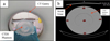 |
Fig. 2 CTDI phantom (a), five ROIs in the CTDI phantom image to evaluate noise and CT number in images (b). |
2.5 Patient study
For image quality assessment, a controlled trial with a parallel group design was conducted. All procedures were in accordance with the ethical standards of ethics committee of Semnan university of Medical Science, Iran. 60 female patients were recruited in this study and divided into two equal intervention and control groups. The Important point is that imaging was parts of the patient’s diagnostic processes and no additional radiation was given to the patients. The criteria for patients to enter the study were female patients aged 20–75 who underwent thoracic CT examinations, and the criteria for patients to leave the study were patients who had a reduced level of consciousness (as a result of trauma, stroke, or other factors) or the patient’s lack of consent to continue participating in the research.
In the CT scan department, after filling informed consent forms by the patients, the optimized shield was placed over the breast organ after the scout scan in the intervention group. In the control group, the patients underwent routine CT imaging of thorax without radiation shield on the breast organ. Before assessing the CT images, image of the shield was removed from the images of the intervention group, and then the images of the intervention and control groups were given to two radiologists. A checklist form was filled about the absence or presence of artifacts (Streak or beam hardening artifact) in the breast, mediastinum, and lung parenchyma regions.
2.6 Statistical analysis
The difference between different groups (the group with different shields and the group without shield) was evaluated using paired t-test. Statistical significance was set at p-value less than 0.05. Furthermore, the Shapiro–Wilk test was used for assessing the normality of the data. All data had a normal distribution.
3 Results
3.1 Breast surface dose on the human thorax phantom
Mean values entrance skin dose (ESD) and percentage of dose reduction in CT imaging without and with radiation shields are listed in Table 1. Using on phantom TLD measurements, the average breast surface dose without radiation shield was 15.03 mGy. The surface dose was reduced (compared to without radiation shield) by 14.17% to 51.69% with the use of radiation shields. The p-values (for comparison of groups with shield compared to the group without shield) of all radiation shields except 100% BaSO4 shield were statistically significant (p < 0.05).
Measured mean breast surface dose during thoracic CT scans with and without the constructed shields.
3.2 Image quality using CTDI phantom
Shields were placed on the CTDI phantom during CT scanning to assess the image quality. Five desirable circular areas (ROIs) were considered in the phantom image with different distances from the shield. The mean values of the pixels inside the ROIs were considered as the average CT number and their standard deviation was considered as the noise. The mean CT number and standard deviation within each ROI were averaged for seven consecutive images. CT numbers shift and image noise increase percentage with and without shields are also shown in Figure 3. The shield with 50% Cu–50% BaSO4 had the lowest destructive effect on the image quality considering the image noise and CT numbers shift. CT number shift and image noise decrease with increasing distance between shield and ROI in the image. The effect of 50% Cu–50% BaSO4 shield on the CT number in all regions except the left peripheral region and on the noise in all regions except the anterior and middle regions was not statistically significant (p > 0.05).
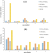 |
Fig. 3 Comparison of CT number shift (a) and noise increase (%) (b) in different ROIs: (1) anterior, (2) posterior, (3) left peripheral, (4) right peripheral, (5) the middle in the CTDI phantom. |
3.3 Image quality analysis by patient study
For the patient study, 50% Cu–50% BaSO4 shield was chosen due to its less impact on image quality. An axial chest CT image of a patient using a 50% Cu–50% BaSO4 shield is shown in Figure 4. Based on the results of the radiologist’s evaluation (Tab. 2), no artifact was observed in the 30 patients of the intervention group. In the case of image quality, there was no significant difference between the intervention and control groups. The radiologists declared that all CT images were had p-value of normal diagnostic quality.
 |
Fig. 4 An example of axial thoracic CT images obtained with 50% Cu-50% BaSO4 shield for shielding of Lung mediastinum (a), for parenchyma (b). |
Data on artifact occurrence in patients with and without 50% BaSO4-50% Cu shield (no artifact, artifact in the lung area, artifact in the mediastinum area, artifact in the breast area).
4 Discussion
The use of CT and its associated radiation dose has been increased in recent years (Bertho et al., 2024). In the recent years, special attention has been focused on more sensitive organs such as the lens of the eye, thyroid, and breast during CT scanning. Although the breast is not the target of the thorax CT examination, the main drawback of this imaging method is that it exposes the breast to ionizing radiation. In large-scale epidemiological studies (Mathews et al., 2013; Miglioretti et al., 2013), it has been shown that the risk of developing cancer in children and young adults is increased due to radiation exposure after CT examinations. Studies on women who survived the atomic bombings of Japan and on patients who underwent multiple radiographs for benign conditions such as tuberculosis and scoliosis have shown the prevalence of cancer in breast tissue after exposure to ionizing radiation (Hoffman et al., 1989; Miller et al., 1989). Therefore, the use of dose reduction techniques for radiosensitive organs is important during CT imaging. IRA and TCM are techniques used to reduce dose in CT scanners. However, these techniques have limitations and are not also existed in all CT scanners (Foley et al., 2013; Lambert et al., 2016). Another method is to use a shield that is placed on the surface organ and reduces the dose to that organ by reducing the radiation intensity in the underlying tissues. In this research, various compositions of Cu and BaSO4 were made in order to reduce the dose for the breast in CT examination. It should be mentioned that for the analysis of dose reduction, in some studies, organ dose and in some other studies, such as this study, the ESD has been reported.
4.1 The effect of new breast radiation shields in dose reduction
Based on data in Table 1, different composition, constructed breast shields reduces the surface dose of breast by 14.77%–51.69%. The difference between the dose reduction of these shields has a maximum of 37.52%. Based on the data presented in Table 3, the efficiency of the constructed bismuth breast shields in this study in reducing the ESD is close to the results of Hopper et al. (2002), Mehnati et al. (2018), Yilmaz et al. (2007), Catuzzo et al. (2010), Huggett et al. (2013), Mendes et al. (2015), Vollmar et al. (2008). These small differences in the results of these studies may be due to differences in imaging techniques, breast sizes and dose measurement tools. The efficacy of the constructed shields in this study suggests that they can be used as dose reduction shields for clinical applications in CT examinations.
Tochaikul et al. (2024) conducted a study to investigate a lead-free radiation shielding material, BaSO4 composite, and evaluated its effectiveness in providing radiation protection. The findings show that the composite containing 30% BaSO4 shows the most effective radiation absorption properties with percentages of 92.15%, 89.26%, and 86.04% in X-ray energies of 40, 60, and 80 kVp, respectively. In addition, composites containing BaSO4 are environmentally friendly and provide good protection against low-dose radiation. Moonkum et al. (2023) performed a study with the aim of investigating a non-lead radiation shield and evaluating its effectiveness in radiation protection. In that study, they studied the characteristics of primary and secondary radiation absorption of shields consisting of silicone rubber, BaSO4 and Bi2O3 in different ratios. The results showed that the protective material with 70% BaSO4 and Bi2O3 has the ability to reduce the radiation dose from 120 kVp X-ray and has absorption properties of 90.19%–94.87% for primary radiation and 92.72%–97.48% for secondary radiation. In addition, silicone rubber shielding materials mixed with BaSO4 and Bi2O3 are environmentally friendly, flexible, and have excellent shielding performance in reducing diagnostic X-ray exposure. However, in the present study, the dose reduction of 100% BaSO4 shield was only 17.14%, which was not statistically significant, and this shield was ultimately rejected in this study.
Comparison of dose reduction due to bismuth shielding between the current study and other relevant studies.
4.2 Effect on noise and CT number shift on the CTDI phantom
The shields were constructed in a rectangular cubic form with different weight percentages of BaSO4 and Cu to find which composition has the least effect on image quality. The shield construction was completely flexible but due to the 2 cm foam which was attached to the shield, the shape of shield seems inflexible (Fig. 2a). 90% Cu–10% BaSO4, 50% Cu–50% BaSO4, 100% BaSO4, 100% Bi shields increased image noise by 4.5%, 9.3%, 8.13%, and 16.5%, respectively and the corresponding CT number shift was 2.43%, 0.77%, 1.10% and 4.00%, respectively. According to the discussed cases, it can be mentioned that scan images with 50% Cu–50% BaSO4 shield, had less noise and CT number shift. The increase in image noise and CT number shift were 2.94 HU and 0.95 HU, respectively, for without and with 50% Cu–50% BaSO4 shield. 50% Cu–50% BaSO4 shield compared to the other compositions, had the lowest degrading effects on the image quality, therefore, this shield was used as the radiation shield material in the patient study for further evaluation. The amount of noise increase and CT number shift in the posterior region was less than the anterior region, and the reason for this is that the posterior region is far from the radiation shield.
In accordance with the literature review, Einstein et al. (2012) demonstrated that the use of commercial Bi shielding of breast during CT coronary angiography increased the noise in the location of the coronary arteries by 2.3 HU. Tappouni et al. (2013) indicated that the use of commercial Bi radiation shield increased the CT number by 20 HU, but the noise reduced to 1.86 HU, which was attributed to the hardening of the beam. The late effect was due to the elimination of low energy rays by bismuth shield. Foley et al. (Foley et al., 2013) demonstrated that when using a Bi shield compared to standard protocols for thorax CT and down-tube flow, the average noise increased from 16.1 HU to 20.4 HU (increase of 4.3 HU). As a result, the use of 50% Cu–50% BaSO4 shield with a distance of 2.52 cm (shield with a thickness of 0.52 cm and a foam pad with a width of 2 cm) protected the breast tissue without degrading the image quality.
4.3 Patient study
By evaluation of the images of the patients in the intervention group and comparing them with the images of the patients in the control group, the effect of 50% Cu–50% BaSO4 shield on the image quality was qualitatively investigated. No artifacts were observed in the lung parenchyma, mediastinum, and breast in the manufactured radiation shields for the breast (Tab. 2). This shield reduced the breast dose without affecting the image quality. Based on literature review, Tappouni et al. (2013) investigated the effect of Bi breast shield and partial CT technique in dose reduction, did not observe any artifacts on image in chest wall, heart and lung parenchyma. The results of the presented study are in agreement with the study by Tappouni et al. (2013).
The use of thorax phantom is the limitation of the present study, for which due to the fact that the thorax phantom and the breast tissue is integral, consisting of only a single piece, the surface dose was measured only on the breast surface. If an anthropomorphic phantom is available, the dose can be measured in different depths of the breast tissue in the presence and absence of shields. Another limitation of this study is the construction of radiation shields in a way that it was not possible to remove low-energy rays on the outer sides of the breast.
5 Conclusions
According to the obtained results, it can be concluded that by using 50% Cu–50% BaSO4 radiation shield for the breast, in addition to dose reduction, the image quality is good in terms of the absence of artifacts, noise and accuracy of the CT number. This shield causes the CT number shift of 0.95 HU and noise increase of 2.94 HU (9.3%), therefore, it can be concluded that this radiation shield can be used for clinical application in thorax CT scanning.
Acknowledgments
The authors would like to thank Cancer Research Center, Semnan University of Medical Sciences, Iran for financial support of this study and also the radiology department of Kosar Hospital (Semnan, Iran) for providing the experimental measurement conditions. The authors would like to extend special thanks from Atomic Energy Organization of Iran (AEOI), Secondary Standard Dosimetry Laboratory (SSDL) for their TLD’s calibration and TLD’s reading services.
Funding
The project was financially supported by Cancer Research Center, Semnan University of Medical Sciences, Iran.
Conflicts of interest
The authors have no conflict of interest.
Data availability statement
The research data associated with this article are included within the article.
Author contribution statement
Marziyeh Behmadi and Mohammad Keshtkar contributed to the study conception and design. Experimental measurements were performed by Farideh Poursoltani, Marziyeh Behmadi, Mahdi Ghorbani and Peyman Hejazi. Data collection and analysis were performed by Farideh Poursoltani, Marziyeh Behmadi and Fatemeh Paknazar. The first draft of the manuscript was written by Farideh Poursoltani, Mohammad Keshtkar, Mahdi Ghorbani and Marziyeh Behmadi and all authors commented on previous versions of the manuscript. All authors read and approved the final manuscript.
Ethics approval
All procedures were in accordance with the ethical standards of Ethics Committee of Semnan University of Medical Sciences with ethical code of IR.SEMUMS.REC.1401.223.
Informed consent
Written informed consent was obtained from all patients.
References
- AAPM Report. 2024. The American Association of Physicists in Medicine (AAPM), AAPM position statement on the use of bismuth shielding for the purpose of dose reduction in CT scanning. Available from: https://www.aapm.org/org/policies/details.asp?type=PP&id=3590 [Last accessed on 17 April 2024]. [Google Scholar]
- Abolhadi S, Parach AA, Mehdipour A, Mehnati P, Sayadi AR. 2023. Evaluation of silicon and 10% bismuth shield with variable thickness compared with constant thickness on the dose reduction and image quality during chest CT examination. Radioprotection 58: 55–60. [CrossRef] [EDP Sciences] [Google Scholar]
- Akhlaghi P, Miri-Hakimabad H, Rafat-Motavalli L. 2014. Effects of shielding the radiosensitive superficial organs of ORNL pediatric phantoms on dose reduction in computed tomography. J Med Phys 39: 238–246. [CrossRef] [PubMed] [Google Scholar]
- Alonso TC, Mourão AP, Santana PC, da Silva TA. 2016. Assessment of breast absorbed doses during thoracic computed tomography scan to evaluate the effectiveness of bismuth shielding. Appl Radiat Isot 117: 55–57. [CrossRef] [PubMed] [Google Scholar]
- Bertho J-M., Bourguignon M. 2024. Un enjeu majeur de radioprotection en imagerie médicale. Radioprotection 59: 1–2. [CrossRef] [EDP Sciences] [Google Scholar]
- Catuzzo P, Aimonetto S, Fanelli G, Marchisio P, Meloni T, Mistretta L, ... Tofani S. 2010. Dose reduction in multislice CT by means of bismuth shields: results of in vivo measurements and computed evaluation. Radiol Med 115: 152–169. [CrossRef] [PubMed] [Google Scholar]
- Colletti PM, Micheli OA, Lee KH. 2013. To shield or not to shield: application of bismuth breast shields. AJR Am J Roentgenol 200: 503–507. [CrossRef] [PubMed] [Google Scholar]
- Curtis JR. 2010. Computed tomography shielding methods: a literature review. Radiol Technol 81: 428–436. [PubMed] [Google Scholar]
- Dere O. 2022. Breast glanduler dose and effectiveness of in-plane bishmut breast shield in routine pediatric female chest CT examination. Int J Curr Med Biol Sci 2: 179–184. [Google Scholar]
- Ehtiati A, Hejazi P, Bakhshandeh M, Arfaee AJ, Bolookat ER, Jadidi M, et al. 2021. The effect of contrast-enhanced computed tomography (CT) scans on the calculated dose of radiotherapy in a thorax phantom. Int J Cancer Manag 14(1):e84158. [Google Scholar]
- Einstein AJ, Elliston CD, Groves DW, Cheng B, Wolff SD, Pearson GD, et al. 2012. Effect of bismuth breast shielding on radiation dose and image quality in coronary CT angiography. J Nucl Cardiol 19: 100–108. [CrossRef] [PubMed] [Google Scholar]
- Feldle P, Grunz JP, Huflage H, Halt D, Schüle S, Sauer L, et al. 2023. Organ-based tube current modulation versus spectral shaping via tin prefiltration: what does bismuth breast shielding add in low-dose lung CT? Eur J Radiol 169: 111157. [CrossRef] [PubMed] [Google Scholar]
- Foley SJ, McEntee MF, Rainford LA. 2013. An evaluation of in-plane shields during thoracic CT. Radiat Prot Dosimetry 155: 439–450. [CrossRef] [PubMed] [Google Scholar]
- Hoffman DA, Lonstein JE, Morin MM, Visscher W, Harris III BS, Boice Jr, JD. 1989. Breast cancer in women with scoliosis exposed to multiple diagnostic X rays. J Natl Cancer Inst 81: 1307–1312. [CrossRef] [PubMed] [Google Scholar]
- Hopper KD. 2002. Orbital, thyroid, and breast superficial radiation shielding for patients undergoing diagnostic CT. Semin Ultrasound CT MR 23: 423–427. [CrossRef] [PubMed] [Google Scholar]
- Huggett J, Mukonoweshuro W, Loader R. 2013. A phantom-based evaluation of three commercially available patient organ shields for computed tomography X-ray examinations in diagnostic radiology. Radiat Prot Dosimetry 155: 161–168. [CrossRef] [PubMed] [Google Scholar]
- Ko CH, Lee SP, Hsieh YC, Lee YH, Yao MM, Chan WP. 2021. Bismuth breast-shield use in chest computed tomography for efficient dose reduction and sufficient image quality. Medicine (Baltimore) 100: e26277. [CrossRef] [PubMed] [Google Scholar]
- Lambert JW, Gould RG. 2016. Evaluation of a net dose-reducing organ-based tube current modulation technique: comparison with standard dose and bismuth-shielded acquisitions. AJR Am J Roentgenol 206: 1233–1240. [CrossRef] [PubMed] [Google Scholar]
- Lestari YM, Anam C, Sutanto H, Arifin Z, Alkian I, Suyudi I, et al. 2023. Evaluation of silicone rubber-lead shield’s effectiveness in protecting the breast during thoracic CT. Biomed Phys Eng Express 9. [PubMed] [Google Scholar]
- Liao YL, Lai NK, Tyan YS, Tsai HY. 2019. Bismuth shield affecting CT image quality and radiation dose in adjacent and distant zones relative to shielding surface: a phantom study. Biomed J 42: 343–351. [CrossRef] [PubMed] [Google Scholar]
- Mathews JD, Forsythe AV, Brady Z, Butler MW, Goergen SK, et al. 2013. Cancer risk in 680,000 people exposed to computed tomography scans in childhood or adolescence: data linkage study of 11 million Australians. Br Med J 346: f2360. [CrossRef] [Google Scholar]
- Mathieu KB, Cody DD. 2013. Partial arc beam filtration: a novel approach to reducing CT breast radiation dose. AJR Am J Roentgenol 200: 601–607. [CrossRef] [PubMed] [Google Scholar]
- Mehnati P, Arash M, Akhlaghi P. 2018. Bismuth-silicon and bismuth-polyurethane composite shields for breast protection in chest computed tomography examinations. J Med Phys 43: 61–65. [CrossRef] [PubMed] [Google Scholar]
- Mehnati P, Arash M, Zakerhamidi M, Ghavami M. 2019. Designing and construction of breast shields using silicone composite of Bismuth for chest CT. Int J Radiat Res 17: 491–496. [Google Scholar]
- Mehnati P, Malekzadeh R, Hussein HA, Obaid NH, Ebrahimiyan S, Sooteh MY, et al. 2023. Trade-off between breast dose and image quality using composite bismuth shields in computed tomography: a phantom study. J Med Imaging Radiat Sci 54: 145–152. [CrossRef] [PubMed] [Google Scholar]
- Mehnati P, Malekzadeh R, Sooteh MY. 2019. New Bismuth composite shield for radiation protection of breast during coronary CT angiography. Iran J Radiol 16: e84763. [CrossRef] [Google Scholar]
- Mendes M, Costa F, Figueira C, Madeira P, Teles P, Vaz P. 2015. Assessment of patient dose reduction by bismuth shielding in CT using measurements, GEANT4 and MCNPX simulations. Radiat Prot Dosimetry 165: 175–181. [CrossRef] [PubMed] [Google Scholar]
- Miglioretti DL, Johnson E, Williams A, Greenlee RT, Weinmann S, Solberg LI, et al. 2013. The use of computed tomography in pediatrics and the associated radiation exposure and estimated cancer risk. JAMA Pediatrics 167: 700–707. [CrossRef] [PubMed] [Google Scholar]
- Miller AB, Howe GR, Sherman GJ, Lindsay JP, Yaffe MJ, Dinner PJ, et al. 1989. Mortality from breast cancer after irradiation during fluoroscopic examinations in patients being treated for tuberculosis. N Engl J Med 321: 1285–1289. [CrossRef] [PubMed] [Google Scholar]
- Moonkum N, Pilapong C, Daowtak K, Tochaikul G. 2023. Evaluation of silicone rubber shielding material composites enriched with BaSO4 and Bi2O3 particles for radiation shielding properties. Mater Res Innov 27: 296–303. [CrossRef] [Google Scholar]
- Parker MS, Kelleher NM, Hoots JA, Chung JK, Fatouros PP, Benedict SH. 2008. Absorbed radiation dose of the female breast during diagnostic multidetector chest CT and dose reduction with a tungsten-antimony composite breast shield: preliminary results. Clin Radiol 63: 278–288. [CrossRef] [PubMed] [Google Scholar]
- Saba V, Keshtkar M. 2019. Targeted radiation energy modulation using Saba shielding reduces breast dose without degrading image quality during thoracic CT examinations. Phys Med 65: 238–246. [CrossRef] [PubMed] [Google Scholar]
- Schöckel L, Jost G, Seidensticker P, Lengsfeld P, Palkowitsch P, Pietsch H. 2020. Developments in X-ray contrast media and the potential impact on computed tomography. Invest Radiol 55: 592–597. [CrossRef] [PubMed] [Google Scholar]
- Tappouni R, Mathers B. 2013. Scan quality and entrance skin dose in thoracic CT: a comparison between bismuth breast shield and posteriorly centered partial CT scans. ISRN Radiol 2013: 457396. [CrossRef] [Google Scholar]
- Tochaikul G, Yokesahachart C, Daowtak K, Pilapong C, Moonkum N. 2024. Preparation and characterization of polylactic acid-based composite incorporating with BaSO4 for low radiation dose shielding. Polym Plast Technol Mater 63: 1733–1744. [Google Scholar]
- Vollmar SV, Kalender WA. 2008. Reduction of dose to the female breast in thoracic CT: a comparison of standard-protocol, bismuth-shielded, partial and tube-current-modulated CT examinations. Eur Radiol 18: 1674–1682. [CrossRef] [PubMed] [Google Scholar]
- Yilmaz MH, Albayram S, Yaşar D, Ozer H, Adaletli I, Selçuk D, et al. 2007. Female breast radiation exposure during thorax multidetector computed tomography and the effectiveness of bismuth breast shield to reduce breast radiation dose. J Comput Assist Tomogr 31: 138–142. [CrossRef] [PubMed] [Google Scholar]
Cite this article as: Poursoltani F, Behmadi M, Keshtkar M, Ghorbani M, Paknazar F, Hejazi P. 2024. Evaluation of barium sulfate-copper breast radiation shield for use in thoracic computed tomography examinations. Radioprotection 59(4): 278–286
All Tables
Measured mean breast surface dose during thoracic CT scans with and without the constructed shields.
Data on artifact occurrence in patients with and without 50% BaSO4-50% Cu shield (no artifact, artifact in the lung area, artifact in the mediastinum area, artifact in the breast area).
Comparison of dose reduction due to bismuth shielding between the current study and other relevant studies.
All Figures
|
Fig. 1 Radiation shield (a), thorax phantom and breast phantom (b), thorax phantom and breast phantom with the shield and dosimetry instrument (c). |
| In the text | |
|
Fig. 2 CTDI phantom (a), five ROIs in the CTDI phantom image to evaluate noise and CT number in images (b). |
| In the text | |
|
Fig. 3 Comparison of CT number shift (a) and noise increase (%) (b) in different ROIs: (1) anterior, (2) posterior, (3) left peripheral, (4) right peripheral, (5) the middle in the CTDI phantom. |
| In the text | |
|
Fig. 4 An example of axial thoracic CT images obtained with 50% Cu-50% BaSO4 shield for shielding of Lung mediastinum (a), for parenchyma (b). |
| In the text | |
Current usage metrics show cumulative count of Article Views (full-text article views including HTML views, PDF and ePub downloads, according to the available data) and Abstracts Views on Vision4Press platform.
Data correspond to usage on the plateform after 2015. The current usage metrics is available 48-96 hours after online publication and is updated daily on week days.
Initial download of the metrics may take a while.