")
")
| Issue |
Radioprotection
Volume 60, Number 1, January-March 2025
|
|
|---|---|---|
| Page(s) | 57 - 64 | |
| DOI | https://doi.org/10.1051/radiopro/2024035 | |
| Published online | 14 March 2025 | |
Article
Thermoluminescence dosimetry in small animal digital radiography, the dose to animals, and their adopters
1
Nuclear Engineering Department, School of Mechanical Engineering, Shiraz University, Shiraz, Iran
2
Radiation Research Center, Shiraz University, Shiraz, Iran
3
Faculty of Engineering, Jahrom University, Jahrom, Iran
* Corresponding author: This email address is being protected from spambots. You need JavaScript enabled to view it.
Received:
25
February
2024
Accepted:
7
August
2024
Abstract
In veterinary radiology, the dose to radiation workers, animals, and the public should be reduced as low as reasonably achievable. Immobilization of the pets is necessary for obtaining images with acceptable image quality. The adopters are usually asked to immobilize their pets during the imaging. Therefore the dose equivalent values received by the animal adopters are also important. This study aims to assess the dose to the hands and whole body of the adopter and the entrance skin dose (ESD) to the animals undergoing radiographic imaging using LiF: Mg, Ti (TLD 100) for one month of work in a veterinary department in Shiraz. The results showed that the dose received by the animals varied from 0.20 mSv to 0.53 mSv. The average dose equivalent values to hands, Hp(0.07), and body, Hp(10) of adopters are 0.35, and 0.14 mSv Respectively.
Key words: Radiation protection / thermoluminescence dosimetry / LiF: Mg / Ti / veterinary medicine / radiographic examination
© E. Saeedian et al., Published by EDP Sciences, 2025
 This is an Open Access article distributed under the terms of the Creative Commons Attribution License (https://creativecommons.org/licenses/by/4.0), which permits unrestricted use, distribution, and reproduction in any medium, provided the original work is properly cited.
1 Introduction
Digital radiology has brought many advantages, such as providing a quick and cost-effective method of obtaining diagnostic images for humans and animals. The modifications made over the last three decades in terms of hardware and software have made digital radiology, an essential diagnostic method for veterinary practitioners. It is becoming increasingly clear to professionals in the veterinary industry that there is a pressing need for clear guidelines and the establishment of best practices to ensure the radiological protection of veterinarians, their staff, and animal adopters. However, the push for this initiative has, so far, primarily focused on the protection of humans rather than the animals themselves, even though the number of veterinary facilities using radiation-based technology has risen significantly in recent years (Owens et al., 2019).
Despite the widespread use of X-ray machines in veterinary medicine, the workload and potential doses received by veterinarians, and their assistants are relatively low (Vatsa et al., 2022; Badawy and Anderson, 2023). Nowadays, there exists the vast majority of data and knowledge about the consequences of ionizing radiation, therefore the radiological protection of human beings has always been of great importance (Fardid et al., 2021; Dietze and Menzel, 1994).
Different imaging techniques are used in modern veterinary medicine, for animals of all shapes and sizes.
Based on the International Commission of Radiological Protection, publication 153, in veterinary imaging, the radiological protection of humans and animals should be considered in the radiological protection program (ICRP 153, 2022). Although imaging modalities provide valuable information, optimization, and radiation risk assessment should also be considered. Like human radiography, there are concerns about the number of repeated radiographs for individual animals. It is essential to increase the awareness of veterinary staff, about the radiation risk, and the optimization of the radiography procedures to reduce the dose to humans, and animals. Research conducted in the US found that most veterinary radiologists and staff believed that doses of ionizing radiation from CT scans did not increase the risk of potentially fatal cancer in the creatures (Gregorich et al., 2018). There is particular concern for breeding animals and those receiving multiple radiographs over their lifetimes, highlighting the importance of being mindful of the potential risks and taking appropriate measures (Gregorich et al., 2018; Lawrence et al., 2015; Hernández-Ruiz et al., 2012; Oh et al., 2018; An et al., 2019).
The immobilization of animals in diagnostic radiology is important for the acquisition of high-quality images. The animals are not capable of remaining still during the imaging, and someone should fix them on the imaging bed.
In veterinary practice, the animal adopters are asked to hold the animals. The International Commission of Radiological Protection, ICRP, recommended that radiation workers in the veterinary facility do not hold animals to avoid repeated exposure. In such cases, the immobilization should be done by people accompanying the patients i.e. their adopters (Mehdizadeh et al., 2019; IAEA, SRS 71, 2013). Therefore they are inevitably exposed to ionizing radiation. According to the international recommendations, and the recommendations of the ICRP, the dose to individual members of the public should be limited (Vano et al., 2015; Dietze and Menzel, 1994; ICRP publication 153; Sousa et al., 2021; Mayer et al., 2018). Therefore the dose received by the animal adopters should be investigated.
This study aims to measure the entrance skin dose, ESD values to the animals, and the dose equivalent values to the body, Hp(10), and hands Hp(0.07) values to the pet adopters during veterinary radiology.
2 Materials and methods
2.1 Digital radiography unit
In the Radiology Department of the School of Veterinary Medicine, Shiraz University, Shiraz, Iran, two X-ray units are employed for performing radiographic examinations on animals, including large and smaller animals. The Philips Practix 100/20 portable X-ray unit (Philips Medical System, Holland) is used for smaller animals such as birds and kittens; while the Philips Super M100 fixed X-ray unit (Philips Medical System, West Germany) is employed for larger animals, such as dogs and cats.
2.2 Preparation of TLD-100 chips
TLD-100 dosimeters, composed of LiF, Mg, and Ti, are widely used in dosimetry, including diagnostic radiology. They are cube-shaped chips measuring 3.1 mm × 3.1 mm × 0.9 mm, commonly used for medical dosimetry (Sadeghi, 2015; Dehghan, 2018; Zehtabian, 2017).
In the first step, the TLD-100 chips were annealed at 400 °C for an hour followed by 80 °C for 20 hours to remove residual signals. Then the element correction coefficient (ECC) for each TLD chip was obtained based on the following equation:
 (1)
(1)
where  is the average of the readings of all dosimeters in a TLD batch, and the TLEi is the reading of the ith TLD and ECCi is the element correction coefficient for the ith TLD chip. Using the ECC values, the reading of the TLDs can be corrected, when used for either personal dosimetry or animal skin dose (ZarifSanayei and Sina, 2024).
2.3 Calibration of TLD chips
The calibration process was performed by using a standard X-ray spectrum. Three calibration curves were drawn, one for entrance skin dose, i.e. ESD, to the animals, and two for personal dose equivalents, i.e. Hp(10), and Hp(0.07), to pet adopters.
2.3.1 Calibration process for ESD measurements for patients (animals)
For calibration, the TLDs were divided into several groups and exposed to various amounts of doses. After exposure, the TLDs were read out using a Harshaw 4500 TLD reader, and the resulting calibration curves were obtained. These curves showed how the TLD response varied with the radiation dose and were used to estimate the entrance surface dose during radiological procedures with similar tube voltages (Sina et al., 2016).
2.3.2 The calibration process for personal dose equivalent for animal adopters
The calibration of the TLDs for personal dose equivalent measurement was performed based on the recommendations of ISO 4037, by using ISO standard phantoms, and by applying the kerma to dose equivalent conversion factors given in the standard above. Two separate calibration curves were drawn for Hp(0.07), and Hp(10). The TLDs were installed on the surface of the ISO slab phantom, and ISO rod phantom, and exposed to known amounts of equivalent dose values, and the calibration curves were prepared.
2.4 Assessment of the ESD for the patient (animal)
To calculate the entrance surface dose (ESD) for pets (dogs, cats, and birds) undergoing X-ray imaging, the annealed TLD chips in thin tissue-equivalent pockets were placed on the surface of their body. The ESD values were measured for 20 animals in the radiology department veterinary medicine school in Shiraz, Iran.
Three TLD chips were placed in a dark plastic pocket and used for measurement of the entrance skin dose (ESD) to each animal (see Fig. 1). Additionally, three TLD chips were used to measure the background level of radiation exposure.
The TLD pockets were installed at the center of the radiation field for each animal. After the imaging procedure was completed, the TLD chips were read out using a TLD reader (Harshaw 4500, USA).
Each TLD chips were read out twice to consider the residual signal. The TLD readings were corrected for the ECC, and the sum of the readings was regarded as the net reading of each chip. The net reading of each chip was corrected for background values.
 (2)
(2)
The corrected reading (nccorrected) of the TLDs for each animal is obtained as follows:
 (3)
(3)
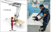 |
Fig. 1 Imaging setup; two small TLD-100 chips were placed on the surface of the animals, and the adopter was standing close to the pet, wearing a protective apron. |
2.5 Equivalent dose to the fingers and body of adopters
As stated in the previous section, the immobilization of the animals during imaging should not be performed by the radiation workers. Therefore, the animal adopters, are asked to hold the animals. Positioning of the animals is often more difficult than the human patients, even children, as they do not follow the orders of the radiographers. Almost all of the images are performed with the assistance of their adopters, as the pets are relaxed in the presence of a familiar individual.
In this section, the dose to the fingers of the adopters holding their pet, Hp(0.07), and the personal dose equivalent to their body, Hp(10), were measured by TLDs. Before beginning the experimental study, the animal adopters were informed and explained about the potential hazards of medical imaging procedures, and written consents were prepared. They were also instructed to use lead aprons with a thickness of 0.5 millimeters of lead available in the hospital. For whole-body dosimetry and obtaining Hp(10) doses, dosimeters were placed under the lead aprons.
Three TLD-100 chips were placed in a plastic badge which has been designed in the form of a card necklace for people’s convenience for measuring Hp(10) to each adopter. For measuring the dose to the hand (finger) of each person, two chips were installed on a ring as shown in Figure 2. The project duration was one month. During the month, 20 animals were imaged. Therefore the dose to 20 adopters were also measured, divided into 6 groups depending on the imaging site.
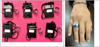 |
Fig. 2 Rings and Cards for measuring Hp(0.07) and Hp (10) respectively. |
3 Results
A total of 20 radiography procedures were performed over a month in the Radiology Department of the School of Veterinary Medicine, Shiraz University, Shiraz, Iran. Over the past decade, there has been a rise in the utilization of radiography in veterinary medicine in Iran. This is a result of people’s growing tendency toward pet adaption. Nevertheless, research on quantifying radiation exposure to animals and monitoring adopter dose equivalent with specific dosimeters is limited.
3.1 Measurement of the entrance skin dose to animals
As previously stated, TLD calibration is required for precise dosimetry. The calibration process utilized five sets of TLDs. Each group received a certain amount of exposure, for obtaining a calibration curve that displayed the dose in µGy versus the TLD reading or thermoluminescence intensity in nano Coulomb (nC) (Fig. 3). The calibration curves can be used for assessment of the entrance skin dose to the animals. Table 1 represents the range, mean value, and standard deviation of the entrance skin dose values measured for different animals undergoing diagnostic imaging using the Philips Practix unit.
 |
Fig. 3 Calibration curve of TLDs used in Animal skin dosimetry. |
ESD estimated for different animals in 20 radiographic examinations
3.2 Results regarding the calibration of dosimeters used for adopters (HP (10) and Hp(0.07))
The calibrations for obtaining the operational quantities in the use of TLDs for absolute dosimetry are displayed as calibration curves in Figures 4 and 5.
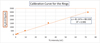 |
Fig. 4 Calibration curve of TLDs used in finger dosimetry (reading of dosimeters based on Hp (0.07)). |
 |
Fig. 5 Calibration curve of TLDs used in body dosimetry (reading of dosimeters based on Hp(10)). |
3.3 Adopters dosimetry results with TLD-100
The TLDs used for the measurement of the adopters were read out using the TLD reader, and the responses were converted to the dose equivalent values using the calibration curves. The imaging is performed for six different sites, i.e. abdomen, cranial thoracic, head, limbs, lung, and neck (see groups 1 to 6 in Tab. 1). Therefore, six groups of adopters were considered for each group. Figures 6 and 7 refer to the average dose received by six different groups of animal adopters.
 |
Fig. 6 Equivalent dose to the hands and fingers of animal adopters calculated in terms of Hp(0.07). |
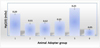 |
Fig. 7 Equivalent dose to the whole body of animal adopters calculated in terms of Hp(10). |
3.4 Uncertainty analysis of the TLD
Table 2 shows the uncertainty analysis for TL dosimeters. As it is obvious, the quadrature combination of type A and B uncertainties are 4%, and 6% respectively. Therefore, the total uncertainty for the measurements is calculated as 7.21%.
Uncertainty budget for thermoluminescence dosimetry.
4 Discussion
The International Commission of Radiological Protection, publication 153, recommends that in veterinary imaging, the main focus should be on reducing the dose to humans, however, animal protection should not be neglected. (ICRP 153, 2022). Based on the publication, in the establishment of radiation protection programs in veterinary departments, the safety of students, or trainers, veterinary staff, radiation protection professionals, and members of the public should be taken into consideration. In the Radiology Department of the School of Veterinary Medicine, Shiraz University, the radiation workers, including radiographers, and veterinarians use appropriate personal dosimeters for dose assessment. In this study, the dose to animals, and the adoptors in the radiological examinations were measured.
Based on the results in the case of animal dosimetry, the average entrance skin dose for all 20 procedures done was 0.36 mGy, with a minimum dose of 0.19 mGy and a maximum dose of 0.53 mGy. In a previous investigation, the dose amount was from 0.62 mGy to the maximum dose of 2.83 mGy (Veneziani et al., 2010). A study examining the dose in the abdomen of 5 animals showed the average dose to be 0.84 mSv, ranging from a minimum of 0.51 mSv to a maximum of 1.87 mSv (Veneziani et al., 2014), while the animals that underwent abdominal imaging in the present study received dose values between 0.20 and 0.53 mSv. Another study showed that the degree of individual exposure among the animals varied greatly, with a range of 0.87 to 2.6 mSv. (Halford and Markham, 1978). A study assessing the dosimetry of mice through the use of TLD (thermoluminescent dosimeter) measurement in combination with micro CT imaging has shown the average radiation dose absorbed by mouse organs to be 760 mSv (Figueroa et al., 2008).
The results of personal dosimetry for the animal adopters showed the average dose to hands, Hp(0.07), was 0.35 mSv and the average dose to the whole body Hp (10) was 0.14 mSv. In radiographic procedures, the dose values to the hand s, Hp(0.07) are higher than the whole-body dose, Hp(10). Because in some cases, the hands of the staff are exposed to the primary radiation.
5 Conclusion
Maintaining radiation safety for veterinarians, their staff, animal adopters, and animals is crucial as the use of ionizing radiation in veterinary diagnostics has significantly increased. The dosimetry of animals and their adopters was performed in veterinary diagnostic radiology. Both dosimetry procedures were performed utilizing TLD-100. The results showed that the dose values measured in this study were lower than the documented quantities in earlier research (Figueroa et al., 2008; Sina et al., 2016; Forrest, 2016; Veneziani et al., 2010). This could be attributed to several factors such as the animals’ thicknesses, and optimized imaging techniques used by the experienced staff of the veterinary department.
In several countries, specific guidelines for radiation safety for veterinarians have been developed. Such cases are those issued by the Australian Radiation Security and Atomic Security Organization (ARPANSA, 2009), and pamphlet by the Heads of the European radiation protection competent authorities (Bly, 2020). Such guidelines are intended only for veterinarians and, to a lesser extent, animal adopters. The guidelines do not address the animals directly. According to the International Commission of Radiological Protection 153, animal protection should not be overlooked in veterinary imaging, but the primary goal should be lowering the dose to humans (ICRP 153, 2022).
Therefore, based on the ALARA principle, the radiation dose to the animal, veterinary staff, animal adopters, and members of the public, should be reduced to as low as reasonably achievable. The optimization of imaging techniques for veterinary imaging, and using protective devices can play a significant role in the radiation protection of people, and animals. To decrease radiation exposure to people and animals, veterinary personnel must be made more aware of the risks associated with radiation and the need to optimize radiography techniques.
Funding
This research did not receive any specific funding.
Conflicts of interest
The authors declare no conflict of interest in regards to this article.
Data availability statement
Due to the nature of the research, no more supporting data is available.
References
- An J et al. 2019. Evaluation of radiation exposure from fluoroscopic examination in small animal veterinary staff using thermoluminescent dosimeters. Veterinární medicína 64(6); 266–270. [Google Scholar]
- Badawy MK, Anderson A. 2023. Radiation protection for comforters and carers in radiology and nuclear medicine. J Medical Radiat Sci 70 (2): 103. [CrossRef] [PubMed] [Google Scholar]
- Bly, R., 2020. Radiation safety of current European practices of therapeutic nuclear medicine: survey results from 20 HERCA countries. [Google Scholar]
- Dietze G, Menzel HG. 1994. Aspects of ICRP 60 and ICRU 47 relevant to individual monitoring of external exposure. Radiat Protect Dosimetry 54 (3-4): 167–173. [CrossRef] [Google Scholar]
- Dehghan, N. and Sina, S., 2020. Measurement of operational dosimetry quantities for nuclear medicine staff. Radiation Protection Dosimetry, 190(2), 119-124. [CrossRef] [MathSciNet] [PubMed] [Google Scholar]
- Faghihi, R., Mehdizadeh, S., Sina, S., Alizadeh, F.N., Zeinali, B., Kamyab, G.R., Aghevlian, S., Khorramdel, H., Namazi, I., Heirani, M. and Moshkriz, M., 2012. Radiation dose to neonates undergoing X-ray imaging in special care baby units in Iran. Radiation protection dosimetry, 150(1), 55-59. [CrossRef] [PubMed] [Google Scholar]
- Fardid R et al. 2021. Evaluation of correlation between DAP (Dose-Area Product) values and cardiologist dose during coronary angiography using Monte Carlo simulation. Iran J Med Phys 18: 306–313. [Google Scholar]
- Figueroa SD et al. 2008. ‘TLD assessment of mouse dosimetry during microCT imaging’. Medical Phys 35: 3866–3874. [CrossRef] [PubMed] [Google Scholar]
- Forrest LJ. 2016. Computed tomography imaging in oncology. Veterin Clin: Small Animal Pract 46: 499–513. [CrossRef] [Google Scholar]
- Gregorich SL et al. 2018. Survey of veterinary specialists regarding their knowledge of radiation safety and the availability of radiation safety training. J Am Veterin Med Assoc 252: 1133–1140. [CrossRef] [PubMed] [Google Scholar]
- Halford DK, Markham OD. 1978. Radiation dosimetry of small mammals inhabiting a liquid radioactive waste disposal area. Ecology 59: 1047–1054. [CrossRef] [Google Scholar]
- Hernández-Ruiz L et al. 2012. Thermoluminescent dosimetry in veterinary diagnostic radiology. Appl Radiat Isotopes 71: 44–47. [CrossRef] [Google Scholar]
- ICRP. 2022. Radiological protection in veterinary practice. ICRP Publication 153. Ann. ICRP 51. [Google Scholar]
- Lawrence YA et al. 2015. Characterization, treatment, and outcome of bacterial cholecystitis and bactibilia in dogs. J Am Veterin Med Assoc 246: 982–989. [CrossRef] [PubMed] [Google Scholar]
- Mehdizadeh, A., Sina, S., Faghihi, R. and Sadeghi, M.H., 2019. An Efficient Radiochemical Method for Extraction of 226Ra From the Soil Samples. Avicenna Journal of Medical Biochemistry, 7(2), 57-60. [CrossRef] [Google Scholar]
- Mayer MN et al. 2018. Use of personal protective equipment in a radiology room at a veterinary teaching hospital. Veterin Radiol Ultrasound 59: 137–146. [CrossRef] [PubMed] [Google Scholar]
- Oh H et al. 2018. Restrainer exposure to scatter radiation in practical small animal radiography measured using thermoluminescent dosimeters. Veterinární Medicína 63: 81–86. [Google Scholar]
- Owens JM et al. 2019. Veterinary radiology—History, purpose, current status and future expectations. Veterin Radiol Ultrasound 60: 358–362. [CrossRef] [PubMed] [Google Scholar]
- Protection, A.R., 2009. ARPANSA-Annual Report Series. [Google Scholar]
- Sadeghi, M., Sina, S. and Faghihi, R., 2015. Investigation of Lif, mg and Ti (TLD-100) reproducibility. Journal of biomedical physics & engineering, 5(4), 217. [PubMed] [Google Scholar]
- Sina S et al. 2016. Evaluation of the entrance skin dose in animals undergoing diagnostic radiology using LiF, Mg, Ti thermoluminescence dosimetry (TLD-100). Iran J Med Phys 13: 118–124. [Google Scholar]
- Sousa CHS et al. 2021. A study to elaborate a technical manual of veterinary radioprotection. J Phys: Conf Ser 12059. [Google Scholar]
- Vano E, Miller DL, Dauer L. 2015. Implications in medical imaging of the new ICRP thresholds for tissue reactions. Ann ICRP 44: 118–128. [CrossRef] [PubMed] [Google Scholar]
- Vatsa MA et al. 2022. Assessment of entrance surface dose (ESD) in some routine X-Rays examinations at the faculty of veterinary medicine Ahmadu Bello University, Zaria. Bayero J Pure Appl Sci 13: 87–91. [Google Scholar]
- Veneziani GR et al. 2010. Thermoluminescence measurements of entrance surface skin dose in exams of dog’s chest in veterinary radiology. Radiat Measur 45: 733–735. [CrossRef] [Google Scholar]
- Veneziani, G.R., Matsushima, L.C. and Campos, L.L., 2014. Evaluation of entrance surface-skin doses in animals submitted on exams of abdomen in veterinary radiology using Tl dosimetry. [Google Scholar]
- Zehtabian, M., Dehghan, N., Danaei Ghazanfarkhani, M., Haghighatafshar, M. and Sina, S., 2017. Measurement of the dose to the family members taking care of thyroid cancer patients undergoing I-131 therapy in nuclear medicine using TLD-100. Radiation protection dosimetry, 174(4), 541-544. [PubMed] [Google Scholar]
- ZarifSanayei A, Sina S. 2024. ‘Measurement of Hp(10), Hp(3) and Hp(0.07) to medical staff in endoscopic retrograde cholangiopancreatography, using thermoluminescence dosimetry’. Radiat Protect Dosimetry ncae008: 473–480. [CrossRef] [PubMed] [Google Scholar]
Cite this article as: Saeedian E, Shakerian M, Zarif Sanayei A, Rakeb Z, Alizadeh FN, Sarshogh S, Sina S. 2025. Thermoluminescence dosimetry in small animal digital radiography, the dose to animals, and their adopters. Radioprotection 60(1): 57–64. https://doi.org/10.1051/radiopro/2024035
All Tables
All Figures
|
Fig. 1 Imaging setup; two small TLD-100 chips were placed on the surface of the animals, and the adopter was standing close to the pet, wearing a protective apron. |
| In the text | |
|
Fig. 2 Rings and Cards for measuring Hp(0.07) and Hp (10) respectively. |
| In the text | |
|
Fig. 3 Calibration curve of TLDs used in Animal skin dosimetry. |
| In the text | |
|
Fig. 4 Calibration curve of TLDs used in finger dosimetry (reading of dosimeters based on Hp (0.07)). |
| In the text | |
|
Fig. 5 Calibration curve of TLDs used in body dosimetry (reading of dosimeters based on Hp(10)). |
| In the text | |
|
Fig. 6 Equivalent dose to the hands and fingers of animal adopters calculated in terms of Hp(0.07). |
| In the text | |
|
Fig. 7 Equivalent dose to the whole body of animal adopters calculated in terms of Hp(10). |
| In the text | |
Current usage metrics show cumulative count of Article Views (full-text article views including HTML views, PDF and ePub downloads, according to the available data) and Abstracts Views on Vision4Press platform.
Data correspond to usage on the plateform after 2015. The current usage metrics is available 48-96 hours after online publication and is updated daily on week days.
Initial download of the metrics may take a while.