")
")
| Issue |
Radioprotection
Volume 60, Number 2, Avril-Juin 2025
|
|
|---|---|---|
| Page(s) | 152 - 158 | |
| DOI | https://doi.org/10.1051/radiopro/2024054 | |
| Published online | 13 juin 2025 | |
Article
Assessment of maternal and fetal radiation exposure during CT pelvimetry in two radiology departments in Morocco
1
Department of Physics, Faculty of Sciences, Ibn Tofail University, Kenitra, Morocco
2
Department of Radiology, Public Hospital, Sidi Slimane, Morocco
3
Department of Radiologic Technology, School of Allied Medical Sciences, Ahvaz Jundishapur University of Medical Sciences, Ahvaz, Iran
4
Al Azhar Oncology Center, Rabat, Morocco
5
Higher Institute of Nursing Professions and Health Techniques, Rabat / Department of Physics, laboratory of materials and subatomic physics, Faculty of Sciences, Ibn Tofail University, Kenitra, Morocco
6
Department of Physics, Faculty of Science, Mohammed V University, Rabat, Morocco
* Corresponding author: Cette adresse e-mail est protégée contre les robots spammeurs. Vous devez activer le JavaScript pour la visualiser.
Received:
22
June
2024
Accepted:
12
November
2024
Abstract
CT pelvimetry scans assess the feasibility of natural childbirth and predict emergency cesarean sections. As the fetus is more radiosensitive than the general population, evaluating and optimizing the associated radiation doses is crucial. Radiology departments require dose data to refine procedures; however, Morocco currently lacks national regulations for dose reference levels (DRLs) specific to CT pelvimetry. Therefore, this study aimed to quantify maternal and fetal radiation doses from CT pelvimetry and establish local DRLs to guide safer imaging practices. A total of 95 pregnant patients who underwent helical scans using 120 kVp voltage and with 30 mAs and 16 mAs across two Moroccan radiology departments, were included in the study. Local DRLs were calculated at the 75th percentile of data, for CTDIvol and DLP, both for individual departments and combined. The average effective dose was calculated with dose conversion factors and estimated using the ImPACT CT Patient Dosimetry Calculator, which also estimated fetal doses in a 70 kg woman. Results revealed statistically significant differences in DRLs between departments (P-values <0.001), with CTDIvol and DLP values recorded at 0.88 mGy and 34.02 mGy · cm, respectively, and inter-departmental DRL discrepancies of approximately 47% for CTDIvol and 35% for DLP. The mean effective dose was 0.47 mSv, while the fetal dose was 1.23 mGy. These findings are pivotal for establishing a foundational framework for DRLs in Morocco aligning with international standards. The study supports radiology departments in benchmarking and optimizing protocols for CT pelvimetry, enhancing patient and fetal safety by minimizing radiation exposure and contributing valuable data for policy development in maternal-fetal radioprotection.
Key words: CT pelvimetry / diagnostic reference levels / effective dose / fetal dose / Moroccan DRLs
© W. Chaanoun et al., Published by EDP Sciences 2025
 This is an Open Access article distributed under the terms of the Creative Commons Attribution License (https://creativecommons.org/licenses/by/4.0), which permits unrestricted use, distribution, and reproduction in any medium, provided the original work is properly cited.
1 Introduction
Over the past few decades, the number of CT examinations in pregnant patients has steadily increased (Lazarus et al., 2009), as has the use of pelvimetric CT to determine the method of delivery (Semghouli et al., 2020; Aabid et al., 2023). Accurate estimation of the dimensions of the birth canal is essential for effective delivery planning and the prevention of complications. Pelvimetry imaging can be conducted before delivery to anticipate the need for an emergency cesarean section. One of the most common indications for an emergency cesarean section is prolonged labor, which aims to prevent fetal distress or suffocation. Additionally, specific obstetric scenarios such as a scarred uterus, a narrow pelvis (Stålberg et al., 2006), breech presentation (Vistad et al., 2013), macrosomia, acquired or congenital deformities of the bony pelvis, or spinal alignment abnormalities can also necessitate a cesarean delivery (Stålberg et al., 2006).
X-ray pelvimetry has evolved significantly with advances in medical imaging technology, since its introduction in the 1940s (Böttcher and Radley, 2001). Conventional X-ray pelvimetry is also widely used to assess the size of the birth canal and it is particularly useful in cases of clinical pelvic stenosis and petite parturient.
CT pelvimetry, on the other hand, offers quicker access and a more comfortable position for pregnant women (Kitai et al., 2020), with simpler measurements and more accurate interpretation. Specifically, a study by Resten et al. (2001) indicated that in CT pelvimetry, the fetus and the mother were exposed to 1/10th of the total dose delivered using conventional X-rays and the dose distribution was more homogenous (Resten et al., 2001). This is while, Phexell et al. (2018) found that the mean absorbed doses to the fetus using various CT pelvimetry methods (topogram, cross-sectional, short spiral, standard spiral, and flash spiral) are higher than those from conventional radiographic techniques; however, the overall dose levels remain low (Phexell et al., 2018). In addition, some studies have suggested magnetic resonance imaging (MRI) (Korhonen et al., 2010), low-dose stereo radiographic imaging (SRI)(Aubry et al., 2018), and ultrasound imaging (Daghighi et al., 2013) for pelvimetry. MRI pelvimetry provides reliable pelvic measurements, but it is also expensive and takes longer to perform, despite its advantages, the use of MRI remains limited by its accessibility (Phexell et al., 2018).
Radiation exposure during pregnancy is a significant concern due to the potential risks it poses to the embryo and fetus, including fetal death, malformations, and developmental delays in cognitive function. Long-term risks are also concerning, such as the potential for hypothyroidism and an increased likelihood of postnatal carcinogenesis (Brent, 2009). Due to the increased radiosensitivity of developing fetal organs, accurately estimating the absorbed dose in the fetus and understanding the associated risks of CT scans are essential (Ray et al., 2010). However, this estimation is challenging, as directly measuring energy deposition in the fetal body is neither feasible nor ethical in humans because of radiation safety and ethical concerns. To address these obstacles, various approaches have been launched to estimate fetal dose, including Monte Carlo simulations using computational anthropomorphic models and physical measurements using phantoms with embedded dosimeters (Xie et al., 2018). Given that the fetus is more radiosensitive than the general population, evaluating and optimizing the associated radiation doses is crucial for patient safety. Dose data are required for radiology departments to refine procedures; however, Morocco currently lacks national regulations for dose reference levels (DRLs) specific to CT pelvimetry. Therefore, this study aimed to quantify maternal and fetal radiation doses from CT pelvimetry and establish local DRLs to guide safer imaging practices. The study findings are pivotal for establishing a foundational framework for DRLs in Morocco aligning with international standards. The study supports radiology departments in benchmarking and optimizing protocols for CT pelvimetry, enhancing patient and fetal safety by minimizing radiation exposure and contributing valuable data for policy development in maternal-fetal radioprotection.
2 Materials and methods
The study adhered to ethical guidelines, ensuring the anonymity of participants and the confidentiality of data.
2.1 Study population and sampling method
The study population consisted of pregnant women referred to the two imaging departments of a public hospital in Morocco for pelvimetric CT scans. The inclusion criteria were pregnant women who underwent pelvimetric CT scans, the existence of the patient’s demographic information, and the scan technical parameters in the related health records. The exclusion criterion was incomplete required information in the medical records or documents. A non-probabilistic purposive sampling method was applied, resulting in a total of 95 patients included in the study. All included pelvimetric CT scans were performed using two General Electric scanners, BrightSpeed 16- slice and LightSpeed VCT 64 slices. Each patient underwent a single helical CT scan as part of the pelvimetry procedure.
2.2 Collected data
Data collection was done retrospectively from patients’ medical records. The collected data included patients’ demographics such as maternal age, height, weight, and gestational age, as well as technical parameters related to the CT machines, including tube voltage (kV), tube current (mA), rotation time(s), pitch, and slice thickness. Table 1 summarizes the technical specifications of the used CT scanners. It should be noted that the acquisition parameters are those of the acquisition protocols, but can be slightly modified for specific patients, leading to differences in the actual doses. These CT machines undergo regular quality control checks to ensure all measured parameters are within an acceptable range.
Applied CT scanners’ characteristics and acquisition parameters.
2.3 Diagnostic reference levels (DRLs)
Diagnostic reference levels (DRLs) introduced in ICRP publication 73 (ICRP, 1996), serve as dosimetric benchmarks for assessing the quality of medical practices and identifying situations that may require corrective measures for routine procedures. These levels should not be exceeded without proper justification. However, these values are not dose limits but guidance values. In CT imaging, as outlined in the Order of May 23, 2019 (ASN, 2019), DRLs are based on the values of the volumetric CT dose index (CTDIvol) and dose-length product (DLP) per acquisition. DRLs are set at the 75th percentile of the median dosimetric values obtained from dose assessments for a specific type of examination (IRSN, 2023). The 75th percentile functions as a threshold beyond which practices may be considered suboptimal, or potentially problematic in terms of dose delivered to the patient (IRSN, 2023). Its international acceptance underscores its importance as a key indicator for DRLs. In our study, the collected data were analyzed first for each department individually and then overall for all departments, to compare doses with international guidelines. Dose parameters were examined and recommended DRLs were established at the 75th percentile of the dose distribution using the statistical package Microsoft Office Excel 2016 to communicate and represent quantitative variables. Statistically significant differences in obtained mean doses were evaluated using the SPSS software version 21.
2.4 Average effective dose
The effective dose (mSv) quantifies the long-term health risks associated with ionizing radiation exposure for the whole population. It can be calculated using the tissue weighting factors recommended by the ICRP (ICRP, 1991) or based on established specific conversion factors (Jessen et al., 1999). Conversion factors were first developed by Shrimpton et al. (1998), using mathematical models (Cristy M., 1987), and were later slightly modified in the European guidelines. Effective dose uses dose conversion factors to provide a metric for managing public exposure and assessing the overall risk to the entire body (Delchambre, 2012). The average effective dose E can be calculated using equation (1):
 (1)
(1)
The conversion factor allows for calculating the average effective dose from the DLP of a CT scan. A conversion factor appropriate for the pelvic region has been applied (Delchambre, 2012). Alternatively, the average effective dose can be estimated using Monte Carlo methods using the ImPACT CT Patient Dosimetry Calculator version 1.0.4 (Fig. 1).
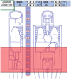 |
Fig. 1 A mathematical phantom with a shaded area covering the pelvic region simulates a CT pelvimetry scan using the ImPACT CT Patient Dosimetry Calculator v1.0.4 to calculate the patient and uterine dose. |
2.5 Fetal dose calculations
The fetal dose was utilized as a proxy for the dose absorbed by the embryo; this was done following the established practice of medical radiation dosimetry. Until September 2006, the dose received by the uterus/fetus was manually calculated based on the technical parameters of the examination. This "manual" method of assessing uterine/fetal dose treats the patient’s pelvis as a 32-cm-diameter plexiglass cylinder and considers the uterus on the axis of this cylinder (Étard and Aubert, 2009). Fetal dose can be calculated using equation (2):
 (2)
(2)
Since 2006, valuations have been carried out using several software packages based on the Monte Carlo method. We estimated the fetal doses for pelvimetric CT using the ImPACT CT Patient Dosimetry Calculator version 1.0.4. This spreadsheet calculates the average effective and organ doses of patients from computed tomography scans. The calculator takes advantage of the National Radiological Protection Board’s (NRPB) Monte Carlo dose data from report SR250, which contains a set of normalized organ dose values for the irradiation of a mathematical phantom, representing a standard 70-kg adult across various CT scanners.
3 Results
The study included data from 95 pregnant women, aged 20 to 40 yr and weighing between 60 and 73 kg, who underwent pelvimetric CT at 36, 37, or 38 weeks of gestation. The average gestational age was 37 weeks. Table 2 indicates the demographics of the included pregnant women in the study. Most studied pelvimetric CT scans (66.31%) were performed at department B while 33.69% of cases were performed at department A. The most common age range among the 95 patients who underwent pelvimetric CT scans was 31–35 yr, representing 42.86% of cases, while the 36–40 age group was the least common, comprising 9.52% of cases. The participants had a mean age of 33 ± 4.65 yr. Most patients (57.23%) weighed between 65 and 69 kg, with an average weight of 69 ± 3.9 kg. Table 3 presents an overview of the variations in CTDIvol, DLP, average effective dose, and fetal dose across each department and both departments combined.
Demographic characteristics of the 95 patients.
Review of the DRLs and doses set for each radiology department.
4 Discussion
The increased use of computed tomography in recent years, including the use of CT pelvimetry to determine the mode of delivery, has raised concerns about patient radiation exposure. In Morocco, this concern is exacerbated by the lack of national legislation and DRLs. To address this issue, this study aimed to evaluate maternal and fetal radiation exposure during CT pelvimetry in two radiology departments in Morocco. To facilitate comparison and discussion of the results of our study with those of other studies, we calculated local DRLs for each department, followed by an aggregate for all departments. These values provide a standardized basis for comparison with the most recent DRLs set internationally in other countries.
Based on the results, of the total 95 included CT scans, one-third of the data were obtained from imaging Department A, and two-thirds were obtained from Department B. At least 30 examinations per radiology department, as was the case with other studies (Barak, 2020; Semghouli et al., 2020) are necessary to comply with the recommendations of the Institute for Radiation Protection and Nuclear Safety (IRSN). This number of pregnant women per department ensures adherence to these guidelines.
Referring to Table 3, in radiology Department A, the local DRLs for CTDIvol and DLP were set at 1.28 mGy and 35.85 mGy-cm, respectively. The calculated average effective dose was 0.49 mSv, which is approximately 2% higher than the estimated average effective dose of 0.48 mSv. Additionally, the estimated fetal dose was 1.5 mGy.
On the other hand, in radiology Department B, the local DRLs for CTDIvol and DLP were 0.87mGy and 26.62 mGy.cm respectively. Both the calculated and estimated average effective doses were 0.43 mSv. The estimated fetal dose for Department B was 0.96 mGy.
The comparison of local DRLs between two radiology departments revealed a statistically significant differences in terms of CTDIvol (mGy) and DLP (mGy · cm) (p-value < 0.001). This indicates that, at the 5% significance level, pregnant patients in Departments A and B do not receive the same doses in terms of CTDIvol and DLP.
Specifically, CTDIvol and DLP values for Department A were approximately 47% and 35% higher than those of Department B, respectively. These differences may be influenced by the technical parameters employed, such as the higher tube current of 60 mA in Department A compared to 40 mA in Department B and the longer tube rotation time of 0.5 s in Department A versus 0.4 s in Department B. While it is essential to consider that radiation dose in CT imaging is influenced by multiple factors, including tube current and exposure time (tube rotation time), and tube voltage (Nagel, 2007; Maldjian and Goldman, 2013), a direct comparison of mA settings between different CT scanner models requires caution due to differences in normalized CTDIw values. This means that the same mA setting may not yield the same dose output across different scanners, leading to potentially misleading conclusions if not properly accounted for (AAPM, 2008; Cody et al., 2010).
However, further efforts are recommended to further optimize and minimize the dose, this can be achieved through comprehensive training of staff in radiation protection and quality control of imaging equipment.
It has also been shown that positioning the patient in a posteroanterior orientation, as opposed to anteroposterior positioning, can result in additional dose reduction during CT scans (Wiesen et al., 1991). Additionally, it is important to adhere to the specified acquisition lengths to optimize dose management. As a viable alternative to CT, the slot-scanning imaging system (EOS) facilitates accurate ex vivo assessment of uterine diameters, provides reliable estimates for obstetric prognosis, and produces measurements comparable to those obtained by CT (Ben Abdennebi et al., 2017). With increasing radiation safety considerations, this low-dose imaging modality has the potential to become the preferred standard for pelvimetry following prospective in vivo validation (Sigmann et al., 2014).
Moreover, the DRLs for all departments for CTDIvol and DLP were set at 0.88 mGy and 34.02 mGy.cm, respectively, as shown in Table 3. Our findings demonstrated significant reductions compared to those of other studies (Tab. 4). In terms of CTDIvol and DLP, respectively, they were approximately 59% and 39% lower than those reported by Semghouli et al. (2020), 2% and 10% lower than those reported by Thibaut et al. (2006), 55% and 39% lower than those reported by Mokubangele et al. (2022), and 12% and 2% lower than those reported by Fayolle et al. (2017).
In addition, as documented in Table 3, the mean effective dose for both radiology departments, assessed as the average of calculated and estimated doses, was 0.47 mSv, significantly lower than values reported in previous studies by Thibaut et al. (2006), Xie et al. (2018), Semghouli et al. (2020), and Mokubangele et al. (2022). This value is below the 1 mSv guideline recommended by the ICRP for managing radiation exposure during pregnancy (ICRP, 2007). It should be noted that this value serves as a guideline for risk assessment rather than an absolute limit for each patient. The mean estimated fetal dose was 1.23 mGy, significantly lower than the fetal doses reported in all previous studies, except for the study by Xie et al. (2018), where our results were approximately 5% higher.
According to international publications, the ICRP sets a threshold of approximately 200 mGy received by the uterus for the appearance of malformations or mental retardation. For the induction of cancer, the risk increases with dose, regardless of the actual dose. The risk of fatal cancer is estimated to be approximately 1% per 100mGy received by the uterus, compared with a natural risk of 0.3% (Étard and Aubert, 2009). The estimated fetal dose in our study was well below the level required to cause deterministic effects in the fetus.
These findings support the results of Osei and Darko Ernest (Osei and Darko, 2013), indicating that CT pelvimetry presents no deterministic risks and poses a minimal likelihood of hereditary radiation effects in the future offspring of the unborn child. The risk of childhood cancer ranged from 1 in 16,000 to 1 in 10,000, while the risk of hereditary disease ranged from 1 in 260,000 to 1 in 106,000, as indicated by Mpeke Mokubangele et al. (2022). These values vary based on acquisition mode, as demonstrated in the study by Benameur et al. (2023), specifically, the estimated risks of childhood cancer were 1 in 16,000 for the sequential mode and 1 in 17,000 for the helical mode. Similarly, the risks of hereditary disease were calculated at 1 in 220,000 for sequential mode and 1 in 270,000 for helical mode.
Although our study provides important contributions, several limitations should be acknowledged. We were unable to include all radiology departments and hospitals conducting CT pelvimetry in Morocco, and our sample of CT pelvimetry exams was limited. For a more comprehensive assessment and to establish robust diagnostic reference levels, a larger patient population and broader inclusion of departments and hospitals are necessary. Another limitation is that the fetal doses in pregnant patients (on average at 37 weeks of gestation) were estimated using the ImpaCT CT Patient Dosimetry Calculator which uses a non-pregnant women phantom. This phantom probably overestimates the fetal dose, which is convenient in the frame of radiation protection.
The DRLs established in our study compared to those from other studies.
5 Conclusion
The maternal and fetal radiation doses from CT pelvimetry across two Moroccan radiology departments were evaluated, with a focus on assessing practices against international dose recommendations. We established local DRLs for each department, revealing that CTDIvol and DLP values for Department A exceeded those of Department B by 47% and 35%, respectively, likely due to differing technical parameters. However, overall DRLs across both radiology departments were lower than reported values in other countries.
Our findings suggest minimal radiation risks for the fetus, with doses well below thresholds associated with childhood cancers and hereditary effects determined in the literature (Mokubangele et al., 2022; Benameur et al., 2023). Without national DRLs in Morocco, our findings highlight the importance of optimizing dose protocols, particularly in Department A, without compromising diagnostic accuracy. Future efforts should emphasize dose management strategies and adherence to best practices in radiation safety.
Acknowledgments
The authors thank the CT radiographers, especially Mr. Farid El Midane and Mrs. Meryem Elhanafi, for their contribution and commitment to this work.
Funding
This research did not receive any specific funding.
Conflicts of interest
The authors declare that they have no conflicts of interest related to this research.
Data availability statement
The research data associated with this article are included within the article.
Author contribution statement
Author W. Chaanoun conceived and designed the study, contributed substantially to its conceptualization and methodology, and drafted the paper. Authors E. Chakir, H. Ouabi, I. Bouadel, M. Talbi, and M. Tahmasbi managed the data analysis of this study, and revised and corrected the manuscript. Authors W.Chaanoun and M. Tahmasbi managed the literature searches, interpreted the results, and carried out the research. All authors read and approved the final version of the manuscript.
Ethics approval
The study adhered to ethical guidelines, ensuring participant anonymity and data confidentiality. Data from patients’ health records were used retrospectively. As calculations were performed using a phantom model, no human subjects were directly involved, and therefore, ethical approval was not required.
Informed consent
This article does not contain any studies involving human subjects.
References
- Aabid M, Semghouli S, Choukri A. 2023. Assessment of computed tomography dose index (CTDI) during CT pelvimetry using monte carlo simulation. Atom Indonesia 49 (1): 2023. https://doi.org/10.55981/aij.2023.1214. [Google Scholar]
- AAPM. 2008. The measurement, Reporting,and Management of adiation Dose in CT, report No 96. [Google Scholar]
- ASN. 2019. Order of 23 May 2019 Approving Decision No. 2019-DC-0667 of the French Nuclear Safety Authority of 18 April 2019 on the Methods for Assessing Ionizing Radiation Doses Delivered to Patients During Radiology Procedures. Official Journal of the French Republic. [Google Scholar]
- Aubry S, Padoin P, Petegnief Y, Vidal C, Riethmuller D, Delabrousse E. 2018. Can three-dimensional pelvimetry using low-dose stereoradiography replace low-dose CT pelvimetry? Diagn Interv Imaging 99 (9): 569–576. https://doi.org/10.1016/j.diii.2018.02.008. [CrossRef] [PubMed] [Google Scholar]
- Barak M. 2020. Dosimetric evaluation of pelviscans in Togo. J Afr d’Imag Méd 12 (3): 133–137. [Google Scholar]
- Ben Abdennebi A, Aubry S, Ounalli L, Fayache MS, Delabrousse E, Petegnief Y. 2017. Comparative dose levels between CT-scanner and slot-scanning device (EOS system) in pregnant women pelvimetry. Phys Med 33 (2016) 77–86 https://doi.org/10.1016/j.ejmp.2016.12.008. [CrossRef] [PubMed] [Google Scholar]
- Benameur Y, Najeh M, Tahiri M, Mkimel M, Baydaoui R. El, Hariri B. El, Mesradi MR, Hilali A, Saad E. 2023. Comparison of foetus organ dose and cancer risks in sequential and helical modes in CT pelvimetry for pregnant patients: a Monte Carlo simulation study. Onkol i Radioter 17 (2): 49–54. [Google Scholar]
- Böttcher B, Radley SC. 2001. Pelvimetry: changing trends and attitudes. J Obstet Gynaecol 21 (5): 459–462. https://doi.org/10.1080/01443610120071983. [CrossRef] [PubMed] [Google Scholar]
- Brent RL. 2009. Saving lives and changing family histories: appropriate counseling of pregnant women and men and women of reproductive age, concerning the risk of diagnostic radiation exposures during and before pregnancy. Am J Obstet Gynecol 200 (1): 4–24. https://doi.org/10.1016/j.ajog.2008.06.032. [CrossRef] [PubMed] [Google Scholar]
- Cody DD, Kim HJ, Cagnon CH, Larke FJ, McNitt-Gray MM, Kruger RL, Flynn MJ, Seibert JA, Judy PF, Wu X. 2010. Normalized CT dose index of the CT scanners used in the national lung screening trial. Am J Roentgenol 194 (6): 1539–1546. https://doi.org/10.2214/AJR.09.3268. [CrossRef] [PubMed] [Google Scholar]
- Cristy MEKF. 1987. Specific Absorbed Fractions of Energy at Various Ages from Internal Photon Sources Oak Ridge National Laboratory. [Google Scholar]
- Daghighi MH, Poureisa M, Ranjkesh M. 2013. Association between obstetric conjugate diameter measured by transabdominal ultrasonography during pregnancy and the type of delivery. Iran J Radiol 10 (3): 185–187. https://doi.org/10.5812/iranjradiol.13191. [CrossRef] [PubMed] [Google Scholar]
- Delchambre M. 2012. Calculation of the cumulative effective dose received by patients in diagnostic CT: creation of a software tool for general practitioners. human medicine and pathology. (dumas-00745007). [Google Scholar]
- Étard C, Aubert B. 2009. Analysis of foetal dose assessed by IRSN from 2004 to 2008. Radioprotection 44 (4): 479–493. https://doi.org/10.1051/radiopro/2009018. [CrossRef] [EDP Sciences] [Google Scholar]
- Fayolle S, Miloudi H, Gonzalez L, Rousselle I, Noel A, Amir S, Dufay F. 2017. A study to establish dose index registry for CT scan examinations. Radioprotection 52 (1): 51–56. https://doi.org/10.1051/radiopro/2017003. [CrossRef] [EDP Sciences] [Google Scholar]
- ICRP Publication 60. 1991. 1990 Recommendations of the international commission on radiological protection. ICRP Ann ICRP 21 (1–3). (Vol. 6) [Google Scholar]
- ICRP Publication 73. 1996. Radiological protection and safety in medicine. Ann ICR P 26. [Google Scholar]
- ICRP Publication 105. 2007. The 2007 recommendations of the international commission on radiological protection. Ann ICRP 37 (Vol. 188) [Google Scholar]
- IRSN. 2023. Analysis of Data for Updating Diagnostic Reference Levels in Radiology and Nuclear Medecine. [Google Scholar]
- Jessen KA, Shrimpton PC, Geleijns J, Panzer W, Tosi G. 1999. Dosimetry for optimisation of patient protection in computed tomography. Appl Radiat Isot 50 (1): 165–172. https://doi.org/10.1016/S0969-8043(98)00024-4. [CrossRef] [PubMed] [Google Scholar]
- Kitai T, Hyodo Y, Morikawa H. 2020. Development of CT pelvimetry using deep learning based reconstruction. Jpn J Radiol Technol 76 (1): 16–25. https://doi.org/10.6009/jjrt.2020_jsrt_76.1.16. [CrossRef] [PubMed] [Google Scholar]
- Korhonen U, Solja R, Laitinenc J, Heinonen S, Taipalea P. 2010. MR pelvimetry measurements, analysis of inter- and intra-observer variation. Eur J Radiol 75 (2): e56–e61. https://doi.org/10.1016/j.ejrad.2009.11.018. [CrossRef] [PubMed] [Google Scholar]
- Lazarus E, DeBenedectis C, North D, Spencer PK, Mayo-Smith WW. 2009. Utilization of imaging in pregnant patients: 10-year review of 5270 examinations in 3285 patients − 1997-2006. Radiology 251 (2): 517–524. https://doi.org/10.1148/radiol.2512080736. [CrossRef] [PubMed] [Google Scholar]
- Maldjian PD, Goldman AR. 2013. Reducing radiation dose in body CT: A primer on dose metrics and key CT technical parameters. Am J Roentgenol 200 (4): 741––747. https://doi.org/10.2214/AJR.12.9768. [CrossRef] [PubMed] [Google Scholar]
- Mokubangele CM, Ebongue AN, Bongue D, Ndontchueng MM, Moifo B. 2022. Evaluation of the fetal-maternal radiation doses in CT-pelvimetry and estimation of the fetal radiation risks in 03 radiology departments in douala-cameroon. Open J Radiol 12 (03): 113–124. https://doi.org/10.4236/ojrad.2022.123013. [CrossRef] [Google Scholar]
- Nagel HD. 2007. CT parameters that influence the radiation dose. In: Radiation Dose from Adult and Pediatric Multidetector Computed Tomography. Medical Radiology (D. Tack, P.A. Gevenois, Eds.). Berlin, Heidelberg: Springer. https://doi.org/10.1007/978-3-540-68575-3_4. [CrossRef] [Google Scholar]
- Osei EK, Darko J. 2013. Foetal radiation dose and risk from diagnostic radiology procedures: a multinational study. Int Sch Res Notices 318425: 7, 2013. https://doi.org/10.5402/2013/318425. [Google Scholar]
- Phexell E, Söderberg M, Bolejko A. 2018. Estimation of foetal radiation dose in a comparative study of pelvimetry with conventional radiography and different computer tomography methods. Int J Radiol Radiat Therapy 5 (4). https://doi.org/10.15406/ijrrt.2018.05.00171. [CrossRef] [Google Scholar]
- Ray JG, Schull MJ, Urquia ML, You JJ, Guttmann A, Vermeulen MJ. 2010. Major radiodiagnostic imaging in pregnancy and the risk of childhood malignancy: a population-based cohort study in ontario. PLoS Med. 2010 Sep 7; 7(9): e1000337. https://doi.org/10.1371/journal.pmed.1000337.PMID:20838660;PMCID:PMC2935460. [Google Scholar]
- Resten A, Mausoléo F, Suissa M, Valéro M, Taylor S, Musset D. 2001. Dosimetry comparison of pelvimetry methods using conventional radiographs and CT. J Radiol 82(9 Pt 1): 991–996. [Google Scholar]
- Semghouli S, Amaoui B, Hakam OK, Choukri A. 2020. Radiation exposure during pelvimetry CT procedures in Ibn Sina Children’s Hospital of Rabat. Radiat Phys Chem 175 (December 2018): 108087. https://doi.org/10.1016/j.radphyschem.2018.12.007. [CrossRef] [Google Scholar]
- Shrimpton PC, Jessen KA, Geleijns J, Panzer W, Tosi G. 1998. Reference doses in computed tomography. Radiat Prot Dosimetry 80 (1-3): 55–59. https://doi.org/10.1093/oxfordjournals.rpd.a032542. [CrossRef] [Google Scholar]
- Sigmann MH, Delabrousse E, Riethmuller D, Runge M, Peyron C, Aubry S. 2014. An evaluation of the EOS X-ray imaging system in pelvimetry. Diagn Int Imaging 95 (9): 833–838. https://doi.org/10.1016/j.diii.2014.01.021. [CrossRef] [Google Scholar]
- Stålberg K, Bodestedt Å, Lyrenäs S, Axelsson O. 2006. A narrow pelvic outlet increases the risk for emergency cesarean section. Acta Obstet Gynecol Scand 85 (7): 821–824. https://doi.org/10.1080/00016340600593521. [CrossRef] [PubMed] [Google Scholar]
- Thibaut C, Marie-Anne O, Douws C, Grenier N, Chateil J. 2006. Interest of MDCT 40-row pelvimetry in dose reduction. J Radiol 87 (10): 1404. https://doi.org/10.1016/S0221-0363(06)87394-4. [CrossRef] [Google Scholar]
- Vistad I, Cvancarova M, Hustad BL, Henriksen T. 2013. Vaginal breech delivery: results of a prospective registration study. BMC Pregnancy Childbirth 13. https://doi.org/10.1186/1471-2393-13-153. [CrossRef] [PubMed] [Google Scholar]
- Wiesen J, Ashmead G, Bellon M. 1991. Improvement in CT pelvimetry. Radiology 178 (1): 259–262. https://doi.org/10.1148/radiology.178.1.1984315.PMID:1984315 [CrossRef] [PubMed] [Google Scholar]
- Xie T, Poletti PA, Platon A, Becker CD, Zaidi H. 2018. Assessment of CT dose to the fetus and pregnant female patient using patient-specific computational models. Eur Radiol 28 (3): 1054–1065. https://doi.org/10.1007/s00330-017-5000-z. [CrossRef] [PubMed] [Google Scholar]
Cite this article as: Chaanoun W, Chakir E, Tahmasbi M, Ouabi H, Talbi M, Bouadel I. 2025. Assessment of maternal and fetal radiation exposure during CT pelvimetry in two radiology departments in Morocco. Radioprotection 60(2): 152–158. https://doi.org/10.1186/1471-2393-13-153
All Tables
All Figures
|
Fig. 1 A mathematical phantom with a shaded area covering the pelvic region simulates a CT pelvimetry scan using the ImPACT CT Patient Dosimetry Calculator v1.0.4 to calculate the patient and uterine dose. |
| In the text | |
Current usage metrics show cumulative count of Article Views (full-text article views including HTML views, PDF and ePub downloads, according to the available data) and Abstracts Views on Vision4Press platform.
Data correspond to usage on the plateform after 2015. The current usage metrics is available 48-96 hours after online publication and is updated daily on week days.
Initial download of the metrics may take a while.